Sunday 29 April 2012

The fragile bone...and its accompanying 'package' of problems 


Elderly over the age of 60 has an increased risk of getting osteoporotic fractures - fractures that occur after a trivial fall that does not normally fracture bones in young and healthy individuals. These fractures frequently affect (starting from the most common) the wrists, the hips and the spine. Fractures involving the spine and hips unfortunately are associated with increased mortality rate of about 10-20 percent, due to immobility after the injury.


Immobilized elderly may suffer from the following problems :


1. Lung infection - lying down for prolonged periods of time will accumulate secretions in the lungs and this relative stasis may cause bacterial infection in the lungs - resulting in breathing difficulty, fever and, if not treated, septicemia (a condition where bacteria spreads to the blood circulation) and ultimately death. This is one of the most serious problem related to prolonged immobility in elderly.


2. Bedsores - pressure on bony prominences for prolonged periods can cause breakage in the skin that, if infected, will lead to more problems. Pressure sores can affect the heel area, the ankles, the lower back (sacral sore) and also the hip region.


3. Deep vein thrombosis. When we walk, the muscles in our legs help to pump blood from the vessels in the legs back to the heart. Immobilized patients will have a relatively 'stagnant' blood in the vessels of their legs, leading to formation of blood clot called thrombus. This accumulation of blood clots in the vessels of the legs is another serious problem. It presents as swelling of the legs, pain in the calves and worse - dislodgment of these clots into the vessels of the chest, a condition called pulmonary embolism, a commonly fatal complication.


4. Urinary tract infection - infection can also affect the urine and its system. This condition may also cause septicemia in immobilized patients.


5. Muscle atrophy (wasting) from disuse. Muscle needs to be used frequently and repeatedly to gain its strength and bulk. Unused muscles will lose its strength causing weakness, loss of normal muscle bulk and contour. The more wasting these patients have, the time needed to redevelop these muscles will be significantly longer.


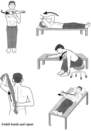
Surgery is normally advised for patients suffering from osteoporotic fractures to avoid all these complications. Nevertheless, after-surgery care is essential to avoid the same set of problems and the aim is to get these individuals mobilized as soon as possible.

Sunday 25 March 2012

Treatment of Frozen Shoulder


   As this condition involves inflammation with subsequent thickening and scarring of the shoulder capsule (the tissue which envelopes the joint), the main problem that most patients face is stiffness of the affected shoulder. Almost all movements will be restricted and their daily activities will be disturbed by this limitation in movement. Overhead activities (wiping the windows, combing hair, playing racket sports) will be difficult and, in the early stage, painful. The patients will also find it difficult to wash or scratch their own back, and strapping bras for ladies will be a nightmare.

   After a careful evaluation and after ruling out other causes, the Doctor will usually start the treatment by thoroughly explaining the condition - it is very important for patients to realize that frozen shoulder is a SELF-LIMITTING condition, meaning that the condition will improve by itself after a period of time even without treatment. Nevertheless, certain situations will need to be treated by surgical means, as will be described later.

   Treatment starts with physical therapy (range of motion exercises). This is the mainstay or the gold standard in all frozen shoulder conditions. Physical therapy ensures that the patient will continue to move the joint despite having a painful shoulder. This is particularly important in the second stage of this condition where the stiffness will be at its maximum and even though the pain is subsiding.

   Passive stretching of the joint in all directions using the opposite limb is done 3-4 times a day, every day. Assistive devices such as a towel, table or wall, and a broom- or walking-stick could also be used to provide maximum effect (see picture).

   Pain can be treated by heat therapy, or analgesics usually prescribed by the treating doctor. This is often unnecessary unless the pain is unbearable and is very much disturbing daily activities. Long-term intake of analgesics is often discouraged.

   In conditions where the stiffness cannot be overcome even with frequent stretching exercises, manipulation of the shoulder (where the surgeon will 'break' the thickened capsular tissue by forced movement under general anesthesia) is an option. Often a surgery is not needed after this procedure, unless the stiffness is persistent. In this rare instance, the surgeon will perform an arthroscopic capsular release whereby the thickened capsule is divided under direct vision (using an arthroscope - see picture below).

An arthroscopic image of the divided capsule (white tissue on the top right area) exposing the underlying muscles (red color).


Please visit your Orthopedic Surgeon for further information on this condition.

Sunday 4 March 2012

Shoulder Pain in a 50-year-old Lady


Madam S is a 50-year-old government servant who came to see me about a year ago with a complaint of right shoulder pain for 3 months' duration. The pain started as vague discomfort in her shoulder whenever she does overhead activities such as wiping her house windows, combing her hair or hanging clothes at home. She also has pain in the shoulder whenever she lies on the affected shoulder at night.

Lately, she thinks that the pain had gotten worse as she also started to have pain even on slight movement in her shoulder while doing her administrative work at the office. She found it difficult to button her bra and also to clean her back while bathing.

Madam S was diagnosed to have diabetes mellitus 3 years ago when routine blood checks revealed that she had elevated fasting blood glucose. She has quite a strong family history of diabetes, with her late father and two brothers were diagnosed to have diabetes.

Examination of her shoulder revealed subtle reduction in movement in her shoulder especially when she was asked to touch her back. Otherwise the examination was unremarkable.

Her X-rays were normal.

“What's wrong with my shoulder Doc?” she asked with a rather concerned face. “Does this condition have something to do with my diabetes? Is it an infection Doc?”

Answer :


This lady was suffering from 'frozen shoulder' - a condition caused by inflammatory shrinking and thickening in the shoulder capsular tissue (the encasing tissue of the shoulder joint).  The inflammation causes the shoulder to become increasingly painful, and, with time, the shoulder becomes stiff and it will be quite difficult to move the affected shoulder in almost all directions. 


The causes of frozen shoulder are :


1. Unknown cause - this condition is also called 'adhesive capsulitis'; in the majority of cases the cause is not identified but there is an association with certain medical conditions such as diabetes mellitus, ischemic heart disease, thyroid gland problems (hyper or hypo-) and Parkinson's disease (a neurological condition that is progressive and characterized by involuntary movements and generalized rigidity of joints). 
   This type of frozen shoulder affects around 2% of the population and typically involves those in the 40-60 years age group. Women are more commonly affected than men.


2. Prolonged immobilization - patients with shoulder injury particularly fractures or dislocations involving or near the shoulder that was operated upon or managed by immobilizing the shoulder joint has a high risk of developing this type of frozen shoulder. 


3. Irradiation to the shoulder region - patients who received radiation therapy (e.g. in breast cancer patients) may also develop this condition.


4. Secondary to a specific shoulder problem for example in shoulder impingement syndrome. Impingement syndrome occurs as a result of tightening of space for the tendons of the shoulder to move, making overhead movements painful and limited. As a result of patients not moving their shoulder due to this pain, the shoulder gets stiffened and a 'secondary' frozen shoulder results.
   A tear in the tendon of shoulder movement (termed rotator cuff tear) may also result in secondary frozen shoulder. 


Stages of Frozen Shoulder 

Frozen shoulder has three stages - these stages tend to overlap and could take as long as two years to resolve even with proper treatment. 


Stage I - 'freezing stage', characterized by increasing pain in the shoulder with movement; however shoulder motion is only mildly affected. This stage lasts from 2 to 4 months.


Stage II - 'frozen stage'. As  the name denotes, the main feature of this stage is stiffness of the shoulder, pain is slowly improving despite worsening in stiffness. The first movement to be affected is inward movement of the arm (for example - difficulty in scratching one's back or strapping the bra), however in advanced stage II all movements are restricted. Duration - 4 months to 1 year.


Stage III - 'thawing stage'. Pain is completely alleviated and stiffness improves during this final stage. This stage takes a year or longer to complete.


** Next week, DocNiz will describe the management options for this condition. Please visit this website again next week
http://shoulderkneedoc.blogspot.com/

Tuesday 21 February 2012

Tips on Reducing Pain in Arthritic Knees


Do you have nagging pain in your knee(s)? Have you injured your knee before from a sports-related mishap or trauma to the knee, and now suffering from its long term effects? Does your doctor told you not to burden your knees but 'forgot' to tell you how? Pain in a damaged knee can be so severe that it might wake the sufferer up from sleep or significantly disturbs his daily activities.

Pain in arthritic knees is caused by either direct rubbing of 'bare bones' against each other or as a result of production of irritant (inflammatory substance) in the joint space, causing pain and swelling in the affected joint.

Arthroscopic pictures of the knee in two patients; one with a normal knee cartilage (above) and another with a degenerated knee (below) where the cartilage has been worn out extensively giving rise to severe pain in the knee with activities.


How do you reduce the burden on your painful knee(s)? Here are some practical tips you can consider :

1. The more weight-bearing you do, the more likely that you increase the burden on your knees. This will in turn cause more pain. Try to reduce unnecessary activities that involves a lot of walking, jumping or running. If certain activities can be done in a non-weight-bearing position such as sitting, do it.
2. If you are overweight, shed some of it. Dietary restrictions coupled with exercises will help you achieve this. Monitoring calorie intake that matches your daily requirement does help. There are a number of activities that can be done to keep fit as well as your weight down without putting excessive burden on your knees (this will be explained further later).
3. Certain positions or activities put a lot of stress on your knees, such as squatting, standing with the knees bent, climbing up and down the stairs, and kneeling on the floor. Utilizing sitting-type toilets will minimize the stress on your knees. Take the elevator or escalator if you have a choice. Use a mop to clean the floor instead of rubbing it using plain cloth, where kneeling is a must.
4. If you want to exercise to keep fit and to reduce weight, perform activities that pose minimal stress on your knees. Swimming and static cycling are two examples of such activities.
5. Use of a walking aid. A walking stick is a social stigma to some people and is considered cumbersome to some, but it is an excellent 'off-loading' device that helps to reduce the load on the knee joint.
6. Do regular 'weight-less' (not weightless) muscle strengthening and joint movement exercises. Static muscle strengthening exercises for the knee (e.g. repeatedly elevating a straight leg off the horizontal plane in a lying position) helps in coordinating and providing support to knee joint movements and helps to reduce pain in the joint. Arthritic joints could easily stiffen from disuse, therefore range-of-motion exercises for the knees are needed to prevent stiffness and contracture.


A straight-leg-raising exercise


For more information, please contact your orthopedic specialist or email to DocNiz at neezlan@yahoo.com


Thursday 9 February 2012

Avoiding Injuries in 'Weekend Warriors'

Injuries can occur from inadequate preparation of muscles and tendons of our body for occasional strenuous activities - the so-called 'weekend warriors'. These injuries can be so disabling that they can either disturb normal weekday activities or completely disrupt them. Worse still, the sufferer may end up admitted to a hospital due to the injury. This will subsequently affect his or her sports activities, productivity and, if prolonged, quality of life.


Futsal is one the common games that may inflict injuries especially among weekend warriors. 


So, what are the common injuries among  these unfortunate 'I-wanna-be-active-but-have-limited-time-for-it' individuals? 


1. Tendinitis - which means inflammation of tendons (the structure which connects muscles to bones). The areas which are prone to this conditions are the heel, elbow, shoulder and knee regions.
2. Sprain - where tissues (such as the muscles or ligaments) are bruised but not torn - most commonly affects the ankle but they may also involve the knee, shoulder and back.
3. Ligament tears - the knee and ankles are most commonly affected by this injury. Complete ligament tears will result in instability in the joints affected, hence will commonly result in permanent pain and instability (sensation that the affected joint 'gives way' during activities) unless they are treated, most commonly through reconstructive surgery where the torn ligament will be replaced by a 'substitute' ligament. Tears may also involve the tendons (e.g. the Achilles tendon at the back of the heel) and meniscus (the 'impact absorption tissue' in the knee).
4. Cartilage injury - cartilage is the lubricating surface in our joints that functions to provide a smooth and congruent movement during activities. Injury to the cartilage can occur during a fall, during an awkward landing from jumping maneuvers, and also from direct hit to the joint (e.g. from a violent tackle during a soccer game). Again, the knees and ankles are the most commonly affected areas. Cartilage injuries are difficult, cumbersome and also expensive to treat, with relatively poor results (compared to other types of injuries) and subsequent sequelae of joint damage and disabling pain.


How do we (as weekend warriors) avoid getting these injuries?

1. Do a proper warm-up and stretching routine before starting any strenuous exercises. These two routines are invaluable in avoiding unexpected injuries. Warm-ups increase the heart rate gradually and thus will lead to steady increase in blood supply to the muscles. Stretching will prepare the muscles for fast, alternating contraction and relaxation that usually occurs during exercises and thus minimizes the risk of muscle sprain and tears.
2. Increase the frequency of exercises that include weekdays as well. As the activities get more frequent and evenly-spaced, the body can withstand more strenuous activities with minimal risk of injuries. 
3. Avoid high-risk sports such as contact sports, high-impact sports (where jumping is done repeatedly), and also pivoting sports where sudden change in directions are needed such as soccer, futsal, basketball and racket games. Swimming and static cycling are two examples of low-risk activities that can be done safely and at the same time allow us to maintain fitness as well as strength.  
4. In situations where high-risk sports cannot be avoided, frequent muscle toning and strengthening exercises will help avoid these injuries. Strong and well-toned surrounding muscles will ensure a more stable joint and thus minimize the risk of injuries to that particular joint.
5. For runners, good shoes with proper arch support and soft heels will help to reduce the impact on the joints of the lower limbs and also protect the ankles from abnormal loading that frequently causes ligament sprains.


Tuesday 24 January 2012


Tips Penjagaan Kaki 
(terutamanya bagi pesakit kencing manis)


Para pesakit kencing manis mempunyai risiko lebih tinggi untuk mendapat jangkitan kuman (yang kadangkala boleh menjadi serius dan mengancam nyawa), terutamanya ke atas anggota badan yang sering terdedah seperti kulit, tangan dan kaki.

Risiko jangkitan ini akan menjadi lebih tinggi sekiranya pesakit-pesakit tersebut mengalami keadaan kebas pada anggota terbabit akibat kencing manis yang tidak terkawal, lantas mendedahkan mereka kepada kecederaan kulit dan anggota yang tidak disedari dan seterusnya mengundang pelbagai bentuk jangkitan sekunder (secondary infection). Lebih menakutkan lagi apabila jangkitan tersebut merebak dan menyebabkan pesakit kehilangan anggota yang terlibat (amputasi, gambar).


Bagaimanakah cara untuk mengelakkan kecederaan dan seterusnya jangkitan pada kaki? Berikut adalah beberapa tips mudah bagi menjaga kaki kita daripada mendapat kecederaan tanpa kita sedari :

  1. Jagalah tahap gula dalam darah anda – ini sangat penting, kerana tahap kencing manis yang tinggi akan menjadikan jangkitan kuman sukar dikawal.
  2. Periksalah kaki anda setiap hari dan awas terhadap kewujudan lepuh-lepuh (blisters), luka (tidak kira berapa kecil saiznya), kemerahan pada kulit, dan tanda bengkak. Gunakan cermin bagi memeriksa kulit pada tapak kaki dan kawasan lain yang terlindung.
  3. Gunakan kasut yang selesa dan sesuai sewaktu berjalan di luar, dan pakailah stokin yang bersih bagi menyerap kelembapan berlebihan pada kulit kaki di dalam kasut. Pakailah kasut yang lebar hujungnya (large toe-box, gambar) bagi mengelakkan jari kaki terkepit di dalam kasut yang akan mengurangkan aliran darah dan menambah risiko kecederaan pada jari kaki. Elakkan penggunaan selipar kerana bahagian kaki akan terdedah kepada benda-benda tajam yang mungkin mencederakan kulit kaki. Jangan berjalan 'kaki-ayam' di luar rumah.

     
    Kasut yang kurang sesuai dipakai (meskipun cantik pada pandangan mata) kerana ruang untuk jari kaki yang sempit


    Kasut ini lebih sesuai kerana ruang jari kaki yang luas dan selesa


  4. Basuhlah kaki setiap hari dengan air yang bersih (!!! jangan gunakan air panas) dan laplah kaki sehingga kering selepas dibasuh. Elakkan dari menggunakan sabuh atau pencuci yang bersifat menghakis (corrosive) kerana ianya akan melukakan kulit kaki. Jangan lupa mengeringkan kulit di antara jari kaki yang mungkin mengundang jangkitan kulat sekiranya dibiarkan lembap selepas dicuci.
  5. Sapulah losyen pada kulit kaki bagi memelihara kelembapan dan mengelakkan rekahan (ini adalah bagi mereka yang mempunyai kulit yang sentiasa kering dan merekah).
  6. Potonglah kuku kaki dengan berhati-hati seminggu sekali dan jangan memotongnya terlalu pendek bagi mengelakkan kecederaan pada lapisan kulit berdekatan kuku. Mintalah pertolongan orang lain sekiranya penglihatan anda tidak begitu jelas. Gunakan pemotong kuku yang sesuai (nail-clippers) dan bukannya pisau bagi mengelakkan kecederaan pada kulit anda sewaktu kuku dipotong.
  7. Elakkan berjalan di kawasan yang terlalu panas (contohnya di kawasan pantai) atau terlalu sejuk (contohnya lantai simen yang sejuk) tanpa alas kaki yang sesuai.
  8. Amalkan senaman regangan kaki dan jari selalu terutama sewaktu berehat di rumah. Ini bagi menggalakkan aliran darah yang lebih berkesan ke anggota-anggota tersebut.
  9. Pergilah berjumpa doktor anda dan jalanilah pemeriksaan kaki secara berkala dengan doktor anda. 

    ** Nota kaki :-

    Untuk mempelajari turutan memotong kuku menurut sunnah Rasulullah SAW - sila ikut panduan rajah ini.


Wednesday 18 January 2012


Shoulder dislocation


A shoulder dislocation may occur as a result of a fall onto an outstretched hand, direct trauma or blow to the shoulder, violent tugging of the arm, electrocution or epileptic fits or secondary to excessive ligamentous laxity of the body. The commonest direction for the dislocation is anterior (where the humeral head comes out to the front of its socket).

Anterior shoulder dislocation is quite common especially in the young and active, and has a high incidence of recurrence (when a dislocation occurs repeatedly it is called instability) - the younger the age of the dislocators, the higher the rate of recurrence. Patients within the age of 20-30 years old has a 80-90% chance of getting repeated episodes of dislocation in the same shoulder.


X-ray of the right shoulder showing a normal shoulder (left) and a dislocated shoulder (below).


Anatomy. The shoulder joint, right picture (medically termed the glenohumeral joint) is essentially an articulation between the humerus (arm bone) and the glenoid (part of the shoulder blade). The size of the humeral articular surface (bigger and rounded in shape) is disproportionate to the size of the smaller and flatter glenoid surface. Without the supporting soft tissues (namely the ligaments and muscles), the shoulder is inherently unstable, just like a golf ball on a tee, (lower left picture). Another stabilizing structure is the labrum, which deepens the glenoid surface (lower right picture).


So why don't we dislocate our shoulder every now and then? It is because of the supporting structures - the ligaments and the muscles that envelope and maintain the humeral head within the glenoid cup which is further deepened by the labrum. When a person dislocates his shoulder, these structures are usually damaged or torn.

Management is initiated by taking a full history, examining the affected shoulder and upper limb (anterior dislocation may injure the axillary nerve which supplies the deltoid muscle - which functions to raise the arm above the shoulder and supplies the sensation at the middle of the shoulder (the so-called 'regimental badge' area), and performing certain maneuvers to reduce the shoulder back into its socket. Reduction must be done as soon as possible as dislocations will distort the blood supply to the humeral head and may cause long-term problems to the patient. Once reduced, the shoulder will be supported in a sling (picture) for 2-4 weeks after which physical therapy will be initiated. The aim of physiotherapy is to strengthen the muscles around the shoulder and to gain back the normal motion in the affected shoulder.

Most patients respond well to conservative treatment. However, patients with persistent instability (either having repeated dislocations or being apprehensive that the shoulder might 'pop-out' during routine activities or those experiencing pain during certain 'vulnerable' positions) may need to undergo surgery to stabilize the shoulder.

For further information, please consult your doctor.